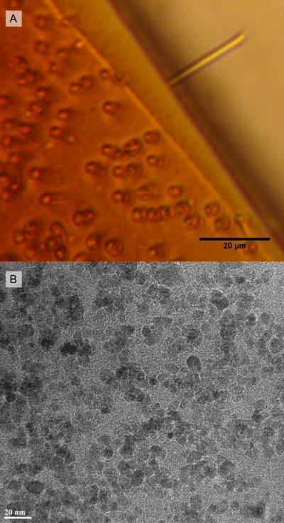
Fig. 2.
A) Cylindrical microstructures (2 μm diam. × 25 μm length) constructed of a 60% FFPDM-SNH2. In the upper-right, one collapsed structure lies parallel to the focal plane and across a crack in the substrate. This microstructure responds to magnetic actuation. No magnetite aggregations are visible at the limits of optical microscopy, indicating microscale homogeneity. B) Transmission electron micrograph of crosslinked 70% FFPDMS-NH2. Magnetite nanoparticles are well-dispersed throughout the material, with diameters ranging from 7 – 10 nm.

