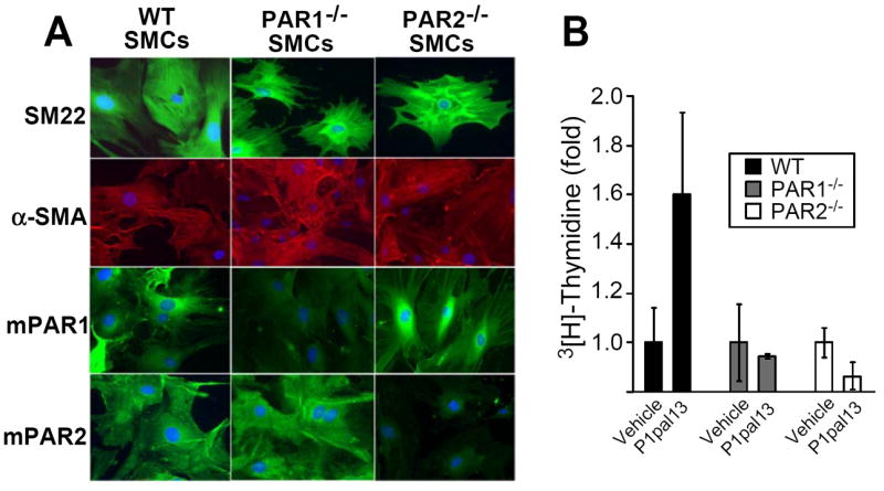
Figure 4. Effect of genetic loss of PAR1 or PAR2 on proliferation.

(A) Primary SMCs were isolated from the carotid arteries of wild-type (WT), PAR1-/- and PAR2-/- mice. SMCs were plated on culture slides and stained with DAPI (blue), or antibodies against SM-22 (top row, green/FITC), α-SMA (second row, red/TRITC), mouse PAR1 (third row, green/FITC), or mouse PAR2 (bottom row, green/FITC). (B) Primary SMCs were subjected to daily treatment of vehicle (0.2% DMSO) or P1pal-13 (3 μM) for 3 d and mitogenesis was measured by incorporation of 3[H]-Thymidine.
