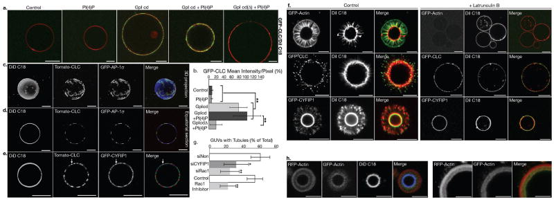
Figure 5. Reconstitution of clathrin-, AP-1-coated carrier biogenesis on model membranes.
(a) DiI C18 labeled giant unilamellar vesicles (GUVs), alone, or containing only PI-4P, only GpI cytoplasmic domains, or both GpI tails and PI-4P, or PI-4P and the GpI cytoplasmic domain devoid of sorting signals (GpIcdΔ), were incubated in the presence of GTP-γ-S with porcine brain cytosol spiked with cytosol of CLC-EGFP expressing cells. They were then imaged by confocal microscopy and (b) CLC-EGFP intensities were determined (n = 3 independent experiments; data represent the mean ± s.d.; pPI4P/no PI4P = 0.386, ≥ 7 GUVs per condition; pGpI/no GpI (no PI4P) = 9.88 × 10⁻⁹, 10GUVs per condition; pGpI/no GpI (with PI4P) = 2.18×10⁻⁷, 7 GUVs per condition; pGpI + PI4P/GpI (no PI4P) = 0.088, ≥ 7 GUVs per condition; pGpI/GpIcdΔ (with PI4P) = 2.5×10⁻²³; ≥ 43 GUVs per condition; Anova single factor analysis). (c–e) DiD C18- labeled GUVs with GpI cytplasmic domains and PI-4P were incubated in the presence of GTP-γ-S and porcine brain cytosol spiked with a mixture of cytosols from cells expressing dTomato-CLC and EGFP-AP-1σ or GFP-CYFIP1. The samples were imaged by confocal microscopy. (f) GUVs with GpI tails and PI-4P were incubated, as in C, with an ATP regenerating system, in the absence (left panels) or the presence of 50 μM Latrunculin B (25 min) (right panels). (g) DiI C18-labeled GUVs with GpI tails and PI-4P were incubated with cytosols of EGFP-actin expressing HEK cells treated with the indicated siRNAs or with 100 nM RAC1 inhibitor (NSC23766). Actin polymerization and tubule formation were analyzed by confocal microscopy, and the number of GUVs displaying EGFP-actin tubes is shown as a percentage of the total DiI C18-positive GUVs (n = 3 independent experiments for siRNA-treated cells and n = 5 independent experiments for Rac1 inhibitor; data represent the mean ± s.d.; > 250 GUVs were analysed per condition; psiCYFIP1 = 0.02, psiRAC1 = 0.0017 compared with control siNon; pRac1 inhibitor = 0.002 compared to control cells; Anova single factor analysis). (h) DiD C18-labeled GUVs with GpI cytoplasmic domains and PI-4P were incubated at 37°C in the presence of GTP-γ-S and porcine brain cytosol spiked with cytosol from RFP-actin expressing HEK cells. After 15 min, cytosol from EGFP-actin expressing cells was added, and the GUVs were incubated for 10 additional min and analyzed by confocal microscopy. Scale bars: 10 μm.

