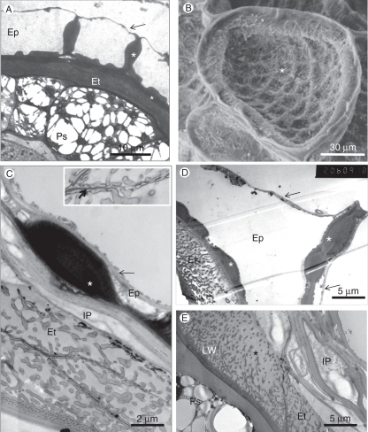
Fig. 8.
The seed coat. (A) TEM image. Cross-section of seed showing a general view of the seed coat and perisperm. In this case, the outer epidermal cell wall is still intact (arrow). Both the radial walls and the inner tangential walls are unevenly thickened (asterisks). The large perisperm cells are filled with oil bodies that surround protein bodies. (B) SEM image of the concave outer surface of a single epidermal cell in which the outer tangential wall collapsed and clings to the inner tangential wall, which has reticulate thickenings (asterisk). (C) The seed coat: an epidermal cell with internal cell wall carrying a large wall thickening (asterisk) to which the external thin cell wall tightly clings (arrow). The endothelium, with diagonal lateral walls, contains many wall protuberances. The appearance of multiple layers is due to the angle of the section. The collapsed inner parenchyma cells sit between these two tissues. Inset: enlarged portion of the cell wall between endothelial cells, showing the dark middle lamella (arrow), traversed by protuberances. (D) Cross-section of the seed coat showing the uneven thickening of the radial wall and the inner tangential walls of an epidermal cell (asterisk). The outer tangential wall of the epidermis collapsed only on one side of the radial wall. The endothelium contains a well-developed labyrinthine wall. (E) The inner layers of the seed coat. In this electron micrograph the collapsed inner parenchyma (IP) is composed of the cell walls of several cells. The endothelium contains labyrinthine wall complex with a density gradient of wall protuberances toward the inner parenchyma. Abbreviations: Ep, epidermis; Et, endothelium; IP, inner parenchyma cell; LW, labyrinthine wall complex; Ps, perisperm.

