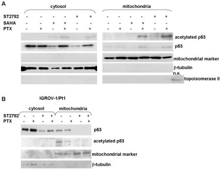
Figure 6. Analysis of mitochondrial p53 translocation induced by ST2782 or SAHA in absence or presence of PTX in IGROV-1 and IGROV-1/Pt1 cells.
The cytosolic and mitochondrial extracts were prepared after 24 h of treatment with PTX alone or combined with HDACi. An antibody against a mitochondrial marker and an antibody against topoisomerase II were used as protein control to ensure a correct subcellular fractionation process. A nuclear extract (n.e.) was used as a positive control for topoisomerase II. A) Comparison of the p53 localization in cytosol or mitochondrial fraction after the combination of PTX (0.12 µM) with ST2782 (10 µM) or with SAHA (3 µM) for 24 h in IGROV-1 cells. One representative experiment of at least three is shown. B) IGROV-1/Pt1 cells were treated for 24 h with PTX (0.012 µM) or ST2782 (10 µM) alone or in combination. One representative experiment of at least three is shown.

