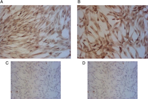
Figure 1.
Immunocytochemical staining of cytokeratin-7 (A) and integrin alpha-1 (B) in primary human EVT cells. Formalin fixed EVTs were stained using the avidin:biotin method. The cells were counterstained with hematoxylin. Positive cells are stained brown and the nuclei are blue. To validate the staining procedure, EVTs were incubated without cytokeratin-7 (C) or integrin alpha-1 (D) primary antibody. Pictures shown were taken with a bright-field microscope at ×10 magnification.

