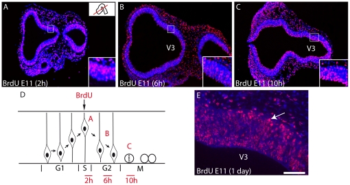
Figure 1. BrdU labeling 2, 6, 10 or 24 h after injection at E11.
Photomicrographs illustrating BrdU labeled nuclei on horizontal sections of the prosencephalon after E11 BrdU injections in the rat. Sections are counterstained with DAPI. Embryos were sacrificed 2, 6 or 10 hours after injection. The distribution of BrdU-labeled nuclei highlights interkinetic movement of these nuclei within the neuroepithelium. (A) Two hours after the BrdU injection, labeled nuclei are external in the neuroepithelium, suggesting that cells are in the S phase. (B) Six hours after injection, labeled nuclei are observed through the thickness of the neuroepithelium, suggesting that these nuclei belong to cells in G2 phase. (C) Finally, ten hours after injection, the ponctiform and intense labeling close to the ventricular surface suggests that mitotic neurons are labeled. (D) Scheme representing the interkinetic movement of nuclei within the neuroepithelium and summarizing the distribution of the BrdU signal as illustrated in A–C. (E) 24 hours after injection, some intensely labeled nuclei are outside of neuroepithelium and invade the dawning mantle layer of dorsal posterior hypothalamus. Scale bar: A–C = 500 µm; E = 100 µm. V3: third ventricle.

