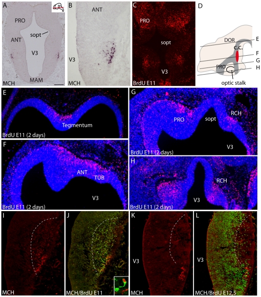
Figure 2. Genesis of MCH neurons.
(A, B) Distribution of preproMCH in situ signal on horizontal sections of an E14 rat embryo. MCH neurons are in a restricted region of the dorsal hypothalamic mantle layer. (C) Distribution of BrdU-labeled nuclei on an E13 rat brain section passing through the ventral hypothalamus after a single BrdU injection at E11. (D–H) Photomicrographs showing BrdU immunohistochemistry on E13 rat horizontal sections counterstained with DAPI, and after BrdU injection at E11; in D, the distribution of BrdU-labeled nuclei (dark grey area) is schematized on a sagittal drawing of the embryonic brain; the red area represent MCH neurons. Sections are arranged from dorsal (E) to ventral (H). BrdU-labeled nuclei are located within a longitudinal region extending from the chiasmatic region to the ventral midbrain, and extending in the preoptic region and ventral telencephalon. (I–L) Distribution of MCH (red) and BrdU-labeled nuclei (green) after multiple injections at E11 (I, J) or E12.5 (K, L). Note the complementary BrdU patterns. Frame in J is a confocal picture of a double labeled MCH/BrdU neuron. For esthetic purpose BrdU is in red and MCH in green. Scale bars: A–C = 500 µm; B, I–L = 250 µm; E–H = 400 µm. ANT: anterior level, hypothalamus; c.c.: ‘cell cord’; MAM: mammillary level, hypothalamus; PRO: preoptic level, hypothalamus; RCH: retrochiasmatic area; sopt: optic sulcus; TUB: tuberal level, hypothalamus; V3: third ventricle.

