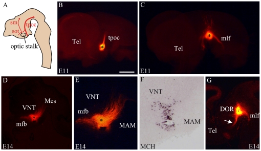
Figure 5. DiI tract tracing from the caudal and dorsal hypothalamus at E11 and E14.
(A) Schematic representation of an embryonic brain sagittal section summarizing the position and direction of the tractus postopticus (tpoc – large red arrow), sot (supraoptic tractus), sm (stria medullaris). (B) A DiI crystal in the caudal hypothalamus of an E11 mouse labels only the tpoc. (C) Control crystal deposited in the ventral mesencephalon of a E11 mouse embryo. The medial longitudinal fascicle (mlf), the tpoc and dorsally directed fibers in the tegmentum are labeled. (D–F) A DiI crystal deposited within the MCH region (J – in situ hybridization for MCH on the same sagittal section) labels caudally but also rostrally directed axons. (G) Control crystals deposited in SN/VTA labeled several tractus as mlf, tpoc, mfb (arrow) or the fasciculus retroflexus. Scale bar: B, C, D, G = 1 mm; E, F = 500 µm. DOR: dorsal thalamus; MAM: mammillary level, hypothalamus; Mes: mesencephalon; Tel: telencephalon; V3: third ventricle; VNT: ventral thalamus.

