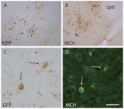
Figure 6. GFP and MCH are expressed by the same cells in MCH-GFP mice.
(A, B) Photomicrographs of two adjacent sections passing through the caudal lateral hypothalamus of an adult mouse brain and labeled by the GFP- or MCH-AS using the standard peroxidase anti-peroxydase procedure. Both AS labeled neurons showing similar distribution patterns in the perifornical region (fx: fornix) and adjacent to the cerebral peduncle (cpd). (C, D): A double immunohistochemical procedure (peroxidase – GFP, immunofluresecnce - MCH), both AS revealed the same neurons. Scale bar: A, B = 500 µm; C, D = 50 µm.

