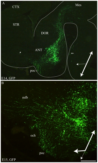
Figure 8. MCH-GFP perikarya and axons at E14–15.
Photomicrographs of the GFP labeling on para-sagittal sections of E14 (A) and E15 (B) MCH-GFP embryos. Caudally directed GFP fibers form a thick tract reaching the mesencephalon. Some of these caudally directed axons are followed ventrally in the neural tube as far as the spinal cord (arrow in A). Another thick bundle of axons run toward the postoptic commisure (poc). Few axons (arrowhead in A) are observed in direction of basal telencephalon at E14 (A). (B) At E15, the number of GFP axons observed in the mfb toward the basal telencephalon dramatically increases. These observations suggest that at E14 most axons follow the tpoc (large white arrow), but some start to take a rostral route (large doted arrow). At E15, it seems that most axons are oriented caudally or rostrally, but less of them take a ventral route. Scale bar: A = 750 µm; B = 200 µm. ANT: anterior level, hypothalamus; CTX: cerebral cortex; DOR: dorsal thalamus; och: optic chiasm; STR: striatum.

