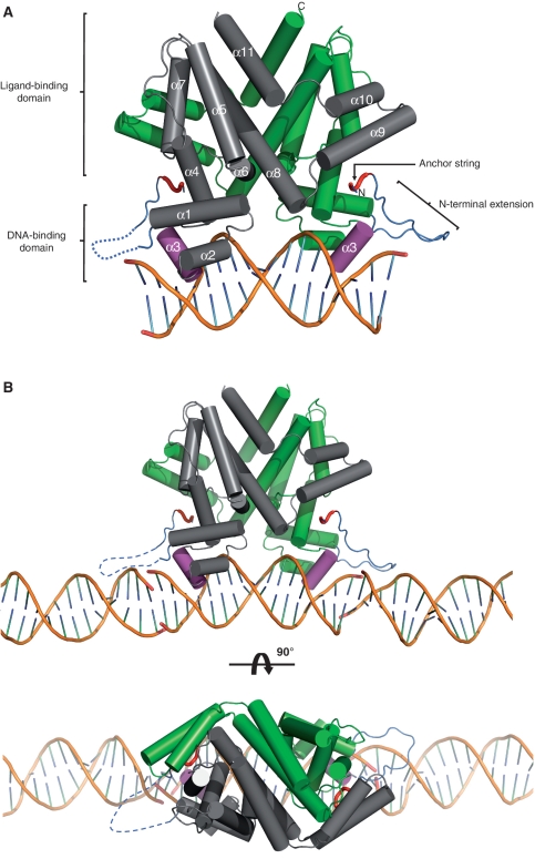
Figure 4.
Structure of the SimR–17-mer complex (A) in isolation or (B) showing the adjacent DNA duplexes in the crystal. A cylindrical helix representation is used to highlight the secondary structure of SimR with key features labelled in (A). One subunit of the biological-relevant dimer is shown in grey and one in green. The recognition helix α3 is shown in magenta, the TFR arm is shown in blue and the N- and C-termini are labelled. The anchor string of the TFR arm (residues 8–11) is shown as a red tube cartoon. The dotted blue line represents the disordered TFR arm in the left-hand SimR subunit. In (B) only the DNA components of the adjacent symmetry complexes are shown in order to highlight the pseudo-continuous DNA filament running through the crystal (See also Supplementary Figure S6 and Figure 7).

