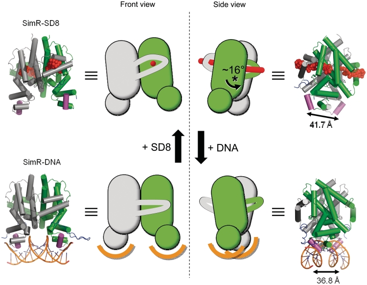
Figure 9.
Structures of SimR–simocyclinone and SimR–DNA together with schematic representations illustrating the rigid-body rotation of the subunits relative to one another. In order to emphasize the subunit rotation, the grey coloured subunits are shown fixed in the same relative orientations. This can be clearly seen in the side view where the green subunit rotates by ~16° relative to the grey subunit; the approximate pivot point is indicated by the asterisk (see also Supplementary Figures S11 and S12). The distances separating the recognition helices α3 and α3′ in the two structures are indicated.

