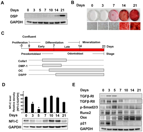
Figure 1. Patterns of gene expression during odontoblast differentiation.
MDPC-23 cells were cultured in differentiation medium for up to 3 weeks. (A) The expression of DSP was evaluated by western blot analysis. GAPDH was used as a loading control. (B) Mineralized nodules stained with alizarin red-S were photographed. (C) Four stages of differentiation were identified: confluent (preodontoblast; 0 days); early odontoblast differentiation (∼7 days); late odontoblast differentiation (7∼14 days); and mineralization (14∼21 days). Solid gray bars indicate the periods of elevated expression of genes indicated during culture. (D) The expression of NFI-C was evaluated by western blot analysis and the results were quantified using ImageJ. (E) TGFβ-RI, TGFβ-RII, p-Smad2/3, Runx2, Osx, and p21 were evaluated by western blot analysis.

