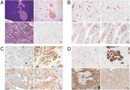
Fig. 1.
PDAC arising in Ptf1a-Cre LSL-KrasG12D p53R172H/+ mice have abundant stroma and sparse vasculature. (A) Normal mouse pancreas (i, ii) and PDAC (iii, iv) stained with H&E (i, iii) and anti-FITC tomato lectin (ii, iv). Perfused FITC tomato lectin identifies functional vasculature during physiologic conditions of assay. (B) Comparison of total vasculature (anti-CD34; i, iii) to functionally perfused vasculature (anti-FITC lectin; ii, iv) within PDAC (i, ii) and duodenum (iii, iv) of a single mouse. Red arrows highlight infrequent CD34+ and anti-FITC lectin positive vessels in adjacent sections of PDAC; in the normal tissue, the two are largely concordant. (C and D) Mouse (C) and human (D) PDAC stained with (i) CD34+ for blood vessels, (ii) cytokeratin to highlight tumor cells, (iii) α-SMA to identify activated myofibroblasts, and (iv) PDGFR-beta. (Magnification: A–C, 50 µm; D. 100 µm.) The lectin perfusion experiment was performed on four untreated mice. All other analyses (CD34, α-SMA, PDGFR-beta, and cytokeratin) were performed on seven untreated or vehicle-treated mice (five vehicle, two no treatment). The selected photomicrographs of invasive tumors immunostained for various antigens are representative of multiple sections of such tumors. Staining pattern among mice within each group showed similar patterns.

