Fig. 2.
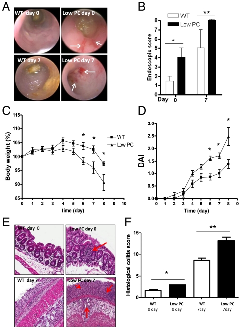
Spontaneous inflammation and increased susceptibility to DSS-induced colitis in low-PC mice. (A) Endoscopic images of mucosal damage in the colon of WT and low-PC mice before (day 0) and after 7 d (day 7) of 2% DSS administration demonstrates the spontaneous mucosal hyperemia and friability in low-PC mice and the presence of mucosal erosions at day 7, as indicated by the white arrows. (B) Quantification of mucosal injury by endoscopic score in WT and low-PC mice before and after treatment. (C and D) Trends toward greater body weight loss (C) and disease activity index (DAI) (D) in low-PC mice compared with their littermates over the course of DSS treatment. (E) Red arrows indicate the presence of small lymphoid follicles at day 0 and increased leukocyte infiltrate at day 7 in the mucosa of low PC mice. (F) Histological damage scoring at day 0 and day 7 in low-PC and WT mice. Histological images were taken using a 20× objective.

