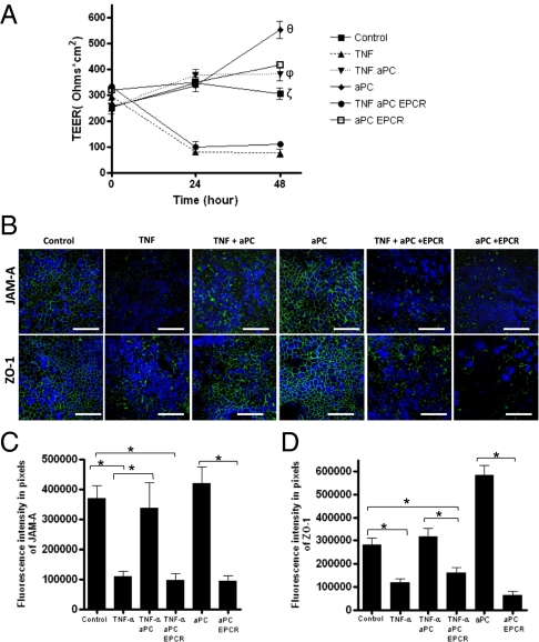
Fig. 4.
Protective effects of aPC on epithelial barrier function are mediated by EPCR. Confluent Caco-2 cells were treated without (control) and with TNF-α (25 ng/mL) in the presence (TNF aPC) or absence of aPC (1 μg/mL), and with (TNF aPC EPCR) or without (aPC EPCR) a 1-h pretreatment with the antibody inhibiting EPCR activity (5 μg/mL). (A) Values of TEER were measured over 48 h of stimulation. (B–D) After 48 h of stimulation, immunofluorescence analysis of JAM-A and ZO-1 (in green) was performed on Caco-2 cells grown on Transwell filters under different conditions: control, TNF, TNF + aPC, aPC, TNF + aPC + EPCR, and aPC + EPCR; DAPI nuclear stain (blue). (Scale bar: 60 μm.) Values are mean ± SEM; the data are representative of three independent experiments. θP < 0.05 (aPC vs. control); ζP < 0.05 (control vs. TNF and TNF aPC EPCR); ϕP < 0.01(aPC vs. TNF and TNF aPC EPCR); *P < 0.01.

