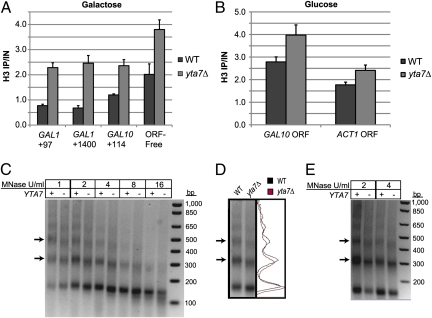
Fig. 5.
Loss of Yta7 resulted in increased nucleosome density. (A) H3 ChIP performed on cells pregrown in YPRaffinose (2%, non-inducing) and then grown with galactose for 2 h. DNA values were quantified by qPCR and are presented as IP/IN for each primer set. (B) H3 ChIP performed on cells growing in YPD. (C) Micrococcal nuclease digestion performed on spheroplasts derived from YPD cultures. Digestions were performed in parallel for 10 min each at 37 °C. Isolated DNA was quantified, and equal amounts were electrophoretically separated on a 2% agarose gel. The arrows indicate the bands corresponding to the di- and trinucleosome products. (D) Overlaid intensity traces of the 2 U/mL MNase lanes presented in C. (E) Micrococcal nuclease digestion, as performed and visualized in C, but performed on spheroplasts derived from YPRaffinose (2%) cultures.

