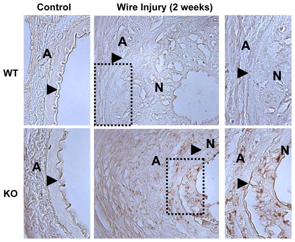
Figure 6. Expression of p21cip1 in mouse femoral arteries.
Sections from uninjured femoral artery (left panels) or 2 weeks after wire injury (middle panels) from WT (upper panels) and KO (lower panels) mice were stained for p21cip1 (in brown). Shown on the right are higher magnification pictures of the dotted areas. The external elastic lamina is indicated by an arrow, N indicates the neointima and A the adventitia. The expression of p21cip1 is evident in femoral arteries from KO mice two weeks after injury.

