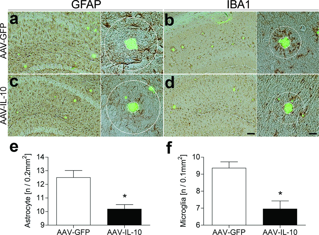
Figure 2. Gene delivery of IL-10 suppresses glial inflammation in APP+PS1 mice.
(a–d) APP+PS1 mice injected with AAV-GFP (a, b) or AAV-IL-10 (c, d) at 3 months of age were sacrificed at 8 months of age. The hippocampal frozen sections were immunostained for GFAP (astrocyte; a, c) or IBA1 (microglia; b, d), and counterstained by TS. Scale bars represent 200µm in low magnification (left, ×40) and 40µm in high magnification (right, ×400). (e, f) Quantification of GFAP (e) or IBA1 (f) positive cells found within the circle surrounding TS-positive Aβ plaques. Radii of outer concentric circles in GFAP-positive cells were 100µm greater than the inner circles that surrounded the compact plaques (a, c), and 50µm greater in IBA1-positive cells (b, d). Bars represent mean ± SEM (n = 5 per group, 10 sections per brain). * denotes P <0.05 as determined by Student’s t-test.

