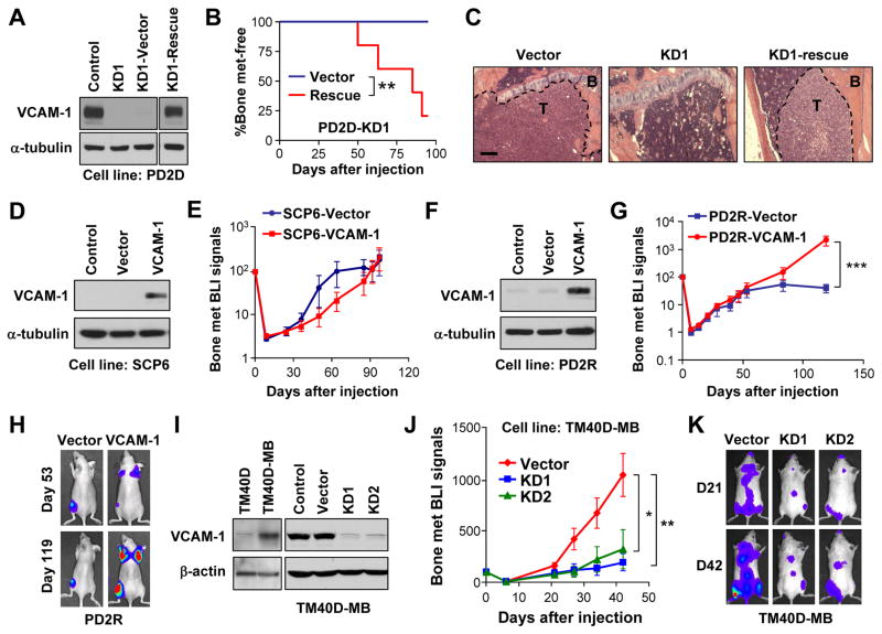
Figure 2. Essential role of VCAM-1 in bone metastasis.
(A) Rescued VCAM-1 expression in the VCAM-1-KD PD2D subline as detected by western blot. (B) Kaplan-Meier representation of bone metastasis relapse by control and VCAM-1-rescued cell lines in (A). N=6. **p < 0.01 by log-rank test. (C) H&E staining of tibia showing presence or absence of overt metastatic tumors (T) in bone (B) by different tumor cells. Scale bar 200μm. (D) Ectopic overexpression of VCAM-1 in SCP6 as detected by western blot. (E) BLI curves of in vivo bone metastasis assay with SCP6 variants. Data represent mean ± SD. N=6. No time point showed statistic significance by Mann-Whitney test. (F) Ectopic overexpression of VCAM-1 in PD2R as detected by western blot. (G) BLI curves of bone metastasis development by control and VCAM-1-overexpressing PD2R cells. Data represent mean ± SEM. N=10. ***p < 0.001 by Mann-Whitney test. (H) Representative BLI of mice in (G). (I) Endogenous expression of VCAM-1 in the murine cell line TM40D and the subline TM40D-MB, and VCAM-1 KD in TM40-MB, as detected by western blot. (J) BLI curves of bone metastasis development by the indicated TM40D-MB cells. Data represent mean ± SEM. N=10. *p < 0.05, **p < 0.01 by Mann-Whitney test. (K) Representative BLI of mice in (J) on Day 21 and Day 42. See also Figure S2.

