
Figure 1. Expression of Presenilins in Sf9 Cells.
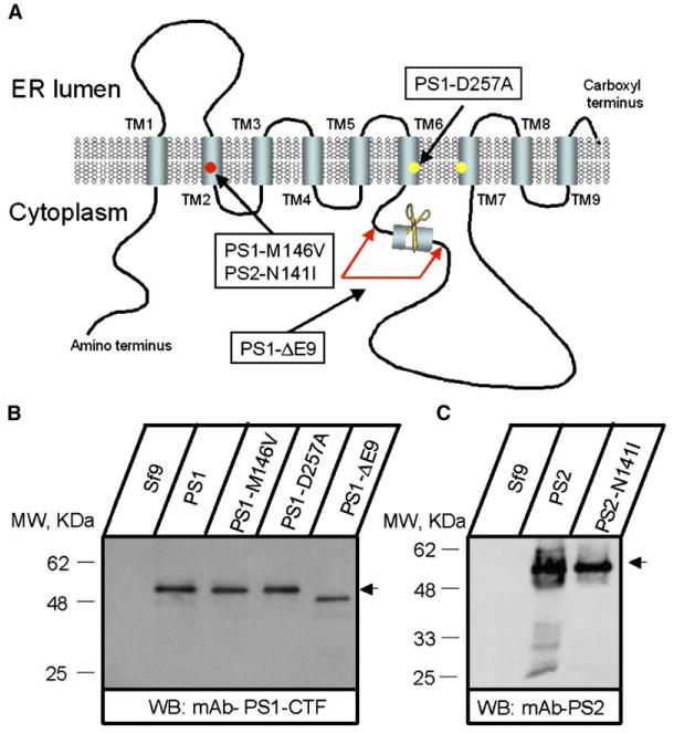
(A) Molecular model of presenilins (based on Laudon et al., 2005). The transmembrane domains (TM1–TM9), locations of aspartate residues critical for γ-secretase activity, and the site of endoproteolytic cleavage are indicated. Positions of PS1-M146V, PS1-ΔE9, PS1-D257A, and PS2-N141I mutations are shown.
(B and C) Expression of PS1, PS2, and mutants in Sf9 cells. Microsomes prepared from noninfected Sf9 cells (Sf9) and from Sf9 cells infected with PS1 and PS2 baculoviruses as indicated were analyzed by Western blotting with anti-PS1 (B) and anti-PS2 (C) monoclonal antibodies.