
Figure 7. Presenilins and ER Ca2+ Homeostasis.
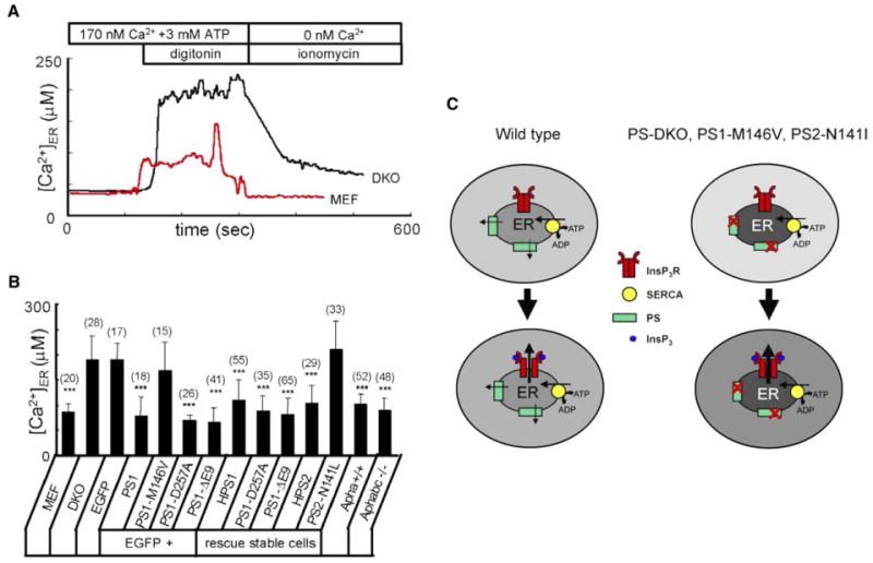
(A) Representative ER Ca2+ traces recorded by ER-loaded Mag-Fura-2 in wild-type (red line) and DKO (black line) MEFs. Cells were loaded with Mag-Fura-2 and permeabilized by 10 μM digitonin in buffer containing 170 nM Ca2+ and 3 mM ATP. At the end of the experiment, the ER membrane was permeabilized with 5 μM ionomycin in the Ca2+-free buffer. 340/380 Mag-Fura-2 ratios were converted to [Ca2+]ER.
(B) The average ER Ca2+ concentration determined as shown in (A) is presented for wild-type MEF cells, DKO cells transiently transfected with EGFP and PS1 rescue constructs, and stably transfected DKO rescue cells as mean ± SD (n = number of cells). When compared to DKO cells transfected with EGFP alone, the ER Ca2+ concentration is significantly (***p < 0.05) lower in DKO cells transfected with EGFP + PS1, EGFP + PS1-D257A, and EGFP + PS1-DE9. The ER Ca2+ concentration is also significantly (***p < 0.05) smaller in HPS1, PS1-D257A, PS1-DE9, and HPS2 stable lines.
(C) Model of Ca2+ homeostasis in wild-type and PS mutant cells. In wild-type cells (left), steady-state ER intraluminal Ca2+ levels are determined by a balance between SERCA pump activity and PS-facilitated passive Ca2+ leak from the ER. In PS-DKO cells or in cells expressing PS1-M146V or PS2- N141I FAD mutants, ER Ca2+ leak function of presenilins is impaired, resulting in higher steady-state intraluminal Ca2+ levels and lower cytosolic Ca2+ levels. Following generation of InsP3 and opening of InsP3R, the amplitude of Ca2+ response is higher in mutant cells (right) than in wild-type cells (left) due to the larger driving force for Ca2+ ions exiting from the ER to the cytoplasm.