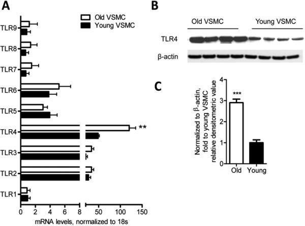
Figure 5. VSMC express increased TLR4 gene expression and protein levels with aging.
TLR gene expression profiles were evaluated in aged and young non-stimulated VSMC by qRT-PCR. TLR4 expression was significantly higher in aged VSMC than young VSMC (A). Western blot analysis showing that aged VSMC exhibit higher TLR4 protein levels than young VSMC (B-C). Results are presented as means ± SEM and in (A) data shown represent one experiment with n = 15 mice as a source of cells / group. In (C) the values are normalized to β-actin expression and represent fold change relative to young VSMC value. In (B) and (C), data shown are representative of two independent experiments, with n = 4 mice / group / experiment as a source of cells. Differences between young and aged VSMC were analyzed using Student's t-test. **, P<0.01, ***, P<0.0001. All comparisons between young and aged groups were paired and performed contemporaneously.

