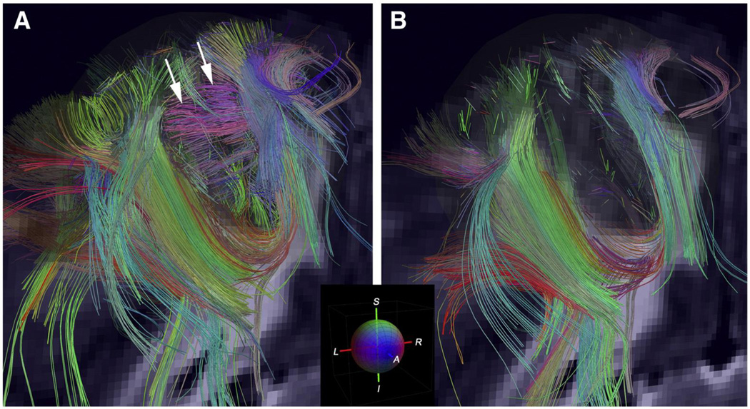
Figure 5.
A = Diffusion spectrum imaging (DSI) tractography of monkey cerebral cortex showing fibers within the white matter of the cortical gyrus, and radiate fibers within adjacent gyri of the cerebral cortex (arrows); B = DTI tractography of a corresponding region shows fewer fibers within the white matter of the gyrus and no radiate fibers within the cortex [175]. Reprinted from Neuroimage, Volume 41/Issue 4, V.J. Wedeen, R.P. Wang, J.D. Schmahmann, T. Benner, W.Y.I. Tseng, G. Dai, D.N. Pandya, P. Hagmann, H. D'Arceuil, & A.J. de Crespigny, Diffusion spectrum magnetic resonance imaging (DSI) tractography of crossing fibers, pp. 1267–1277, Copyright 2008, with permission from Elsevier.

