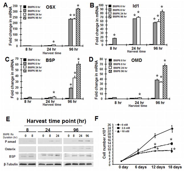
Figure 2. BMP6 upregulates Osterix and other osteoblast genes dependent upon treatment duration.
hMSC were differentiated into osteoblasts in serum-free osteogenic medium and in the presence or absence of BMP6 (20 nM) for 8, 24 and 96 hours, respectively. Real-time PCR analysis of the relative expression of the transcription factors (A) Osterix and (B) Id1 and the extracellular matrix proteins (C) bone sialoprotien (BSP) and (D) osteomodulin (OMD). The data represent the mean values ±SD of triplicate experiments normalized to the housekeeping gene β-actin. (F): BMP6 treatment decreases hMSC proliferation. hMSCs (2×10⁴ per well in 6-well plates) were cultured in the assay medium with 20% FBS in the presence of BMP6 at 0, 2.5 and 10 nM. Cumulative cell number was counted at 6, 12 and 18 days. Statistical differences (*) of p < 0.05 between the BMP6 treated and untreated cell lines were determined using t-test.

