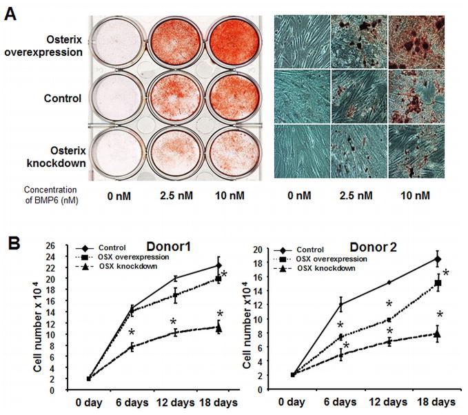
Figure 5. Osterix regulates hMSC osteoblastogenesis and proliferation.
(A) Left panel: Osterix-overexpressing and knockdown hMSC were treated with BMP6 for 96 hours at 0, 2.5 and 10 nM, and stained with Alizarin red S to evaluate calcium phosphate mineral, compare to the control cells. Right panel: Alizarin red S stained wells visualized using bright-field microscopy. (B): Transduced hMSC from donor 1 and donor 2 were cultured in assay medium with 20% FBS (growth conditions). Cumulative cell number was counted at 6, 12 and 18 days. Statistical differences (*) of p< .05 between the Osterix overexpression or knockdown and control PRLP2 cell lines were determined using t-test.

