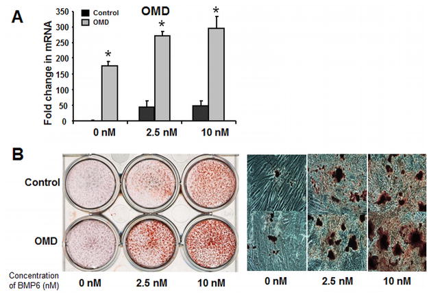
Figure 6. Enforced expression of osteomodulin promotes BMP6 induced hMSC osteoblastogenesis.
(A): Real-time quantitative PCR analysis to determine OMD expression level in the PRLP2-OMD retroviral supernatant treated hMSC in osteogenic medium with BMP6 for 4 days at 0, 2.5 and 10 nM. (B): Left panel: OMD-overexpressing hMSC were treated with BMP6 for 96 hours at 0, 2.5 and 10 nM, and stained with Alizarin Red S to evaluate calcium phosphate mineral, compared to the control cells. Right panel: OMD-overexpressing hMSC were stained with Alizarin Red S to evaluate mineralization and visualized using bright-field microscopy at 14th day. The Q-PCR data represent the mean values ±SD of triplicate experiments normalized to the housekeeping gene β-actin. Statistical differences (*) of p < .05 between the Osterix knockdown and control PRLP2 cell lines were determined using t test.

