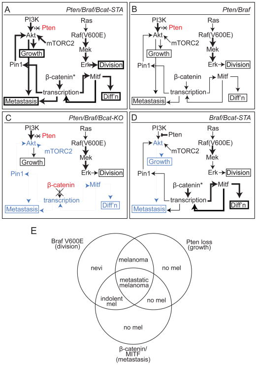
Figure 8. Signaling and phenotypic overview of murine melanoma models.
(A) Pten/Braf/Bcat-STA melanomas are characterized by rapid growth, high degree of melanocytic differentiation, and extensive metastasis. These phenotypes are associated with increased MAPK/Erk and PI3K/Akt signaling as well as elevated Mitf and Pin1 levels. Bold text: relatively increased levels and/or activity compared to other models, blue shading: relatively decreased compared to other models, red shading: genetically inactivated, *: stabilized.
(B) Pten/Braf melanomas exhibit rapid tumor growth, but only intermediate melanocytic differentiation and metastasis. Signaling pathway activation, Mitf, and Pin1 are intermediate in this model.
(C) Pten/Braf/Bcat-KO melanomas exhibit tumor growth, but significantly reduced melanocytic differentiation and almost no metastasis. Inactivation of β-catenin in this model is associated with reduced PI3K/Akt signaling and Mitf levels.
(D) Braf/Bcat-STA melanomas are characterized by long latency and slow tumor growth, as well as lack of metastases. These tumors are differentiated and heavily pigmented due to high Mitf levels, but show reduced PI3K/Akt pathway activation.
(E) A three-pathway synergy is observed when MAPK/Erk, PI3K/Akt, and β-catenin/Mitf signaling pathways are simultaneously activated in melanoma. If any of these three changes is not present, the metastatic melanoma phenotype is abrogated.

