
Figure 4.
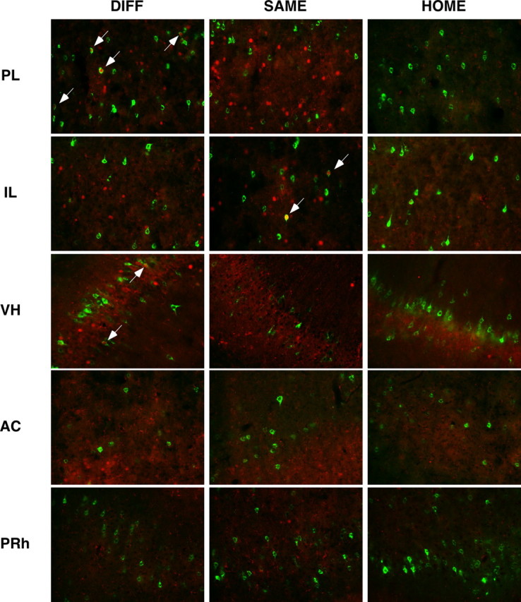
Photomicrographs of representative double-labeled neurons in the PL, IL, VH, AC, and PRh for each behavioral group (DIFF, SAME, HOME). White arrows indicate double-labeled neurons; CTb-positive neurons are stained green, and c-fos-positive neurons are stained red.