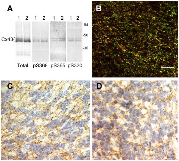
Fig. 1. Brain shows phosphorylation at S365, S325/328/330 and S368.
(A) For SDS-PAGE and Western blot, two mouse brains were quickly removed and frozen in liquid nitrogen. The tissue was pulverized in the presence of sample buffer containing protease and phosphatase inhibitors to prevent dephosphorylation during tissue homogenization. Samples from brain 1 and 2 (one in each lane) were run on 10% PAGE, blotted and probed with NT1 antibody to total Cx43 or antibodies to Cx43 phosphorylated at S368 (pS368), S365 (pS365) and S325/328/330 (pS330) [73]. (B) Immunofluorescence of OCT embedded frozen sections of brain using total Cx43 (Cx43IF1 [72] – in green) and S325/328/330 ([78] - in red) antibodies. Immunohistochemistry of formalin-fixed, paraffin-embedded tissue sections stained with Hematoxylin/Eosin showing cerebellar cells staining at opposed plasma membranes (brown) for total Cx43 (C) and phospho-S368 (D). Phospho-S368 levels appeared to be highest in the cerebellar regions of the brain. Bars are 20 μm.

