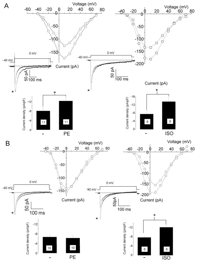
Figure 4.
A dominant negative mutant of PKD1 inhibits α-adrenergic enhancement of L-type Ca2+ currents in neonatal rat cardiomyocytes. The cells were cultured on glass bottom dishes and L-type Ca2+ currents were measured by patch-clamp. Membrane potential was held at −40 mV to avoid contamination by T-type Ca2+ currents. Current–voltage (IV) relationship before (square) and 10 min after addition of 20 μM PE (left) or 1 μM ISO (right) measured on a particular cardiomyocyte in control condition (A) or infected with a GFP-tagged PKD1 dominant negative mutant adenovirus (B). Insets are traces obtained upon depolarization from −40 mV to 0 mV before and after PE (left,*) or ISO (right,*) addition. Lower bar graph of the current density (pA/pF) with mean and standard error of the mean (s.e.m.) measured at 0 mV. Number of measured cells is indicated in the bar graph. *P < 0.02.

