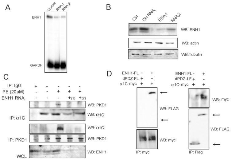
Figure 5.
ENH1 siRNA inhibits PKD1 interaction with α1C in neonatal rat cardiomyocytes. Cells were infected 48 h with the ENH1-RNAi adenovirus. Then total RNA and proteins were extracted. (A) WB analyses for ENH1, actin, and tubulin as control (Ctrl), under the unrelated siRNA (Ctrl RNAi), ENH1 RNAi (RNAi1, RNAi2) conditions. (B) Detection of ENH1 and GAPDH RNA messenger by RPA from control, RNAi1, and RNAi2 conditions. ENH1 siRNA efficiency was tested independently by WB (n = 3) or RPA (n = 3). (C) Protein extracts from control or infected with ENH1 RNAi adenovirus conditions were immunoprecipitated using an anti-PKD1 antibody or an anti-α1C antibody (n = 3). Precipitates were analysed by WB. The lowest panel, WB shows ENH1 expression. (D) Flag-tagged ENH1 (ENH1-FL) and PDZ deletion mutant ENH1 (dPDZ-FL) were co-transfected with myc-tagged α1C (α1C-myc) in 293T cells. ENH1 or PDZ deletion mutants-Fl or α1C-myc were immunoprecipitated from cell extracts using an anti-Flag (IP:Flag) or an anti-myc (IP:myc) monoclonal antibodies. Immunoprecipitates were analysed by WB using monoclonal HRP conjugated anti-Flag (WB:Flag) or anti-myc antibodies (WB:myc).

