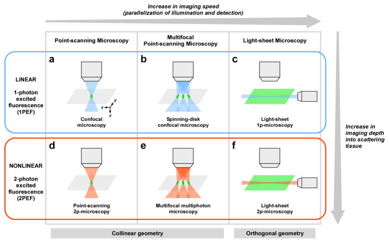
Figure 1. Strategies for improving acquisition speed in current fluorescence microscopy techniques.
Similar strategies have been developed in linear (a-c) and nonlinear (d-f) microscopy. Both point-scanning (a and d) and multifocal (b and e) approaches use a collinear geometry: the illumination and the detection paths are collinear. Light-sheet microscopy (c and f) uses an orthogonal geometry: the illumination path is orthogonal to the detection path. Pixel rate range typically from 105–106 pixels.s−1 in point-scanning microscopy to 107–108 pixels.s−1 in light-sheet microscopy. While nonlinear microscopy generally provides deeper imaging than linear microscopy, differences exist in imaging depth performance between the different implementations of nonlinear microscopy, as discussed in the text.

