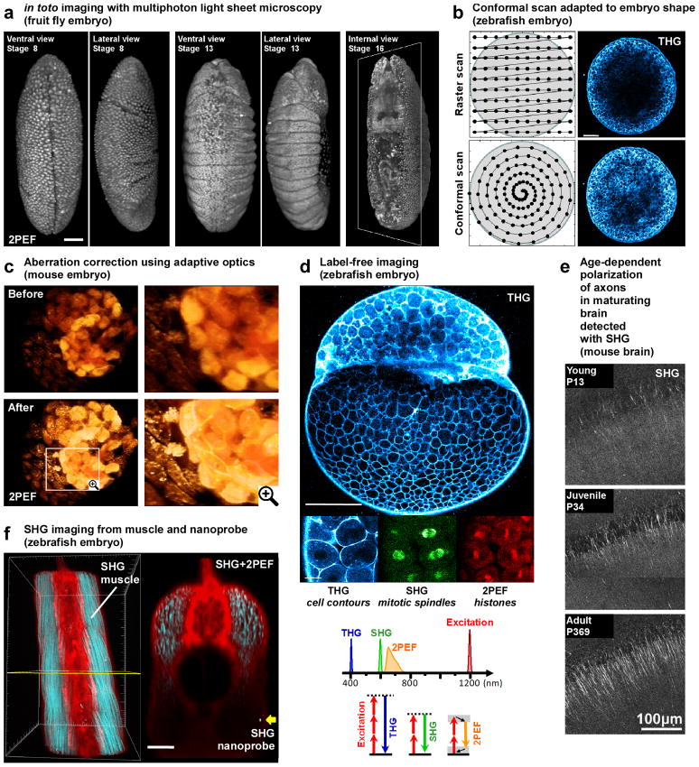
Figure 2. Selected applications of advanced multiphoton microscopy in developmental biology.
This figure illustrates various recent applications of multiphoton microscopy in developmental biology. (a) Long term imaging of Drosophila embryos using light-sheet 2p-microscopy (reprinted from [16], scale bar is 50 μm). (b) Standard raster scanning in multiphoton microscopy produces inhomogeneous signal levels across the embryo. Conformal scanning adapted to the embryo shape provide homogeneous signal for the entire image (image adapted from [30] and reprinted with permission from AAAS, scale bar is 100 μm). (c) 3D rendering of point-scanning 2p-microscopy imaging of a mouse embryo before and after correction of sample-induced aberrations showing improvement in both signal intensity and spatial resolution (adapted from [33] and reprinted with permission from OSA). (d) Simultaneous 2PEF, SHG and THG imaging of zebrafish embryos allows detection of histones, mitotic spindles and cell contours, respectively. Such label-free imaging with SHG and THG signals has been used for 3D cell segmentation and tracking and for reconstructing the cell lineage of early zebrafish development (image adapted from [30] and reprinted with permission from AAAS, scale bars are 200 μm (top) and 20 μm (bottom)). (e) Maturation of axons and dendrites is probed with SHG in mouse hippocampal slices (adapted from [49], copyright (2008) National Academy of Sciences, USA). (f) 3D reconstruction of combined 2PEF (from Bodipy TR dye in red) and SHG (from muscle endogenous signal and BaTiO3 nanoparticle in blue) signals recorded in zebrafish embryos: the strong SHG signal from the nanoprobe (yellow arrow) is detected twice deeper than the endogeneous SHG from muscles (image adapted from [51] and reprinted with permission from authors, scale bar is 50 μm).

