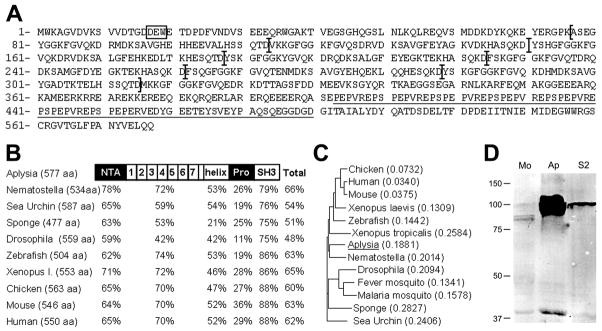
Figure 1. Cortactin is expressed in Aplysia nervous system.
A: Deduced amino acid sequence of Aplysia cortactin. It contains the Arp2/3 binding motif DEW (box), seven and a half tandem repeats (delimited by brackets) followed by a helix, a proline-rich region (underlined), and an SH3 domain. B: Domain-based and full-length cortactin amino-acid sequence comparison between Aplysia and other species (Nematostella: GI # 156408902; Sea Urchin: 47551203; Sponge: 37951188; Drosophila: 24648611; Zebrafish: 51870938; Xenopus laevis: 148222482; Chicken: 45382633; Mouse: 75677414; Human: 20357552). The percentages of sequence homology are indicated. C: Phylogenetic tree built from full length cortactin sequence from several species (same sequences as in B plus Xenopus tropicalis: 58332478; Fever mosquito: 157131030; Malaria mosquito: 158293853) using Vector NTI software. D: Cortactin Western blotting of mouse brain (Mo), Aplysia nervous system (Ap) and Drosophila S2 cell line (S2) extracts. Twenty micrograms of total protein was loaded for each tissue. The doublet in the Aplysia tissue could be clearly seen following shorter exposure. Molecular weights of standards (in kilo daltons) are indicated on the left.

