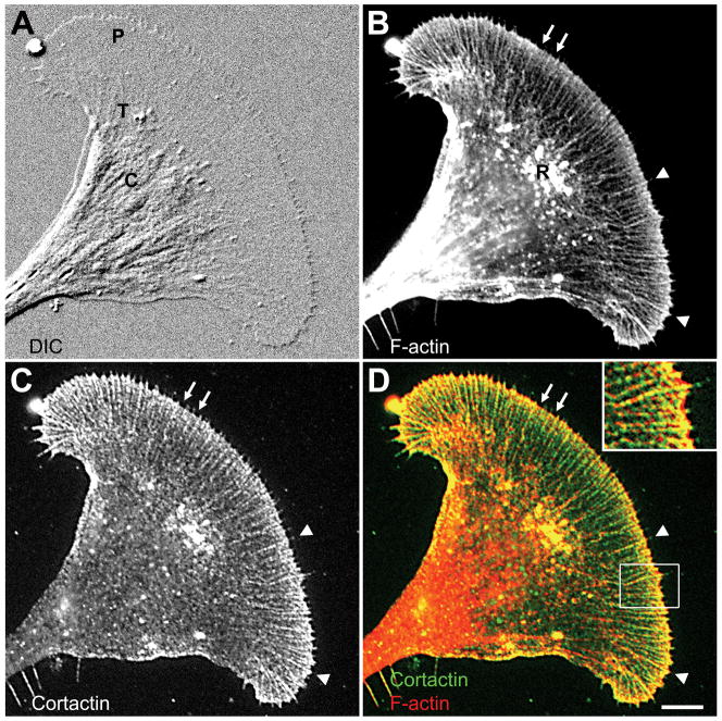
Figure 2. Cortactin colocalizes with F-actin in Aplysia growth cones.
Cortactin and F-actin show a strong colocalization in the growth cone peripheral domain (P) and transition zone (T) when compared to the central domain (C). A: DIC image of a fixed growth cone. B: F-actin staining of the same growth cone using Alexa 488 phalloidin revealed filopodial bundles (arrowheads), lamellipodial veils at the leading edge (arrows) and T zone ruffles (R). C: Cortactin staining with 4F11. D: Overlay of F-actin and cortactin staining. Inset: zoom of the boxed region showing strong actin/cortactin colocalization in filopodia and the lamellipodia at the leading edge. Bar: 10 μm (5 μm for inset).

