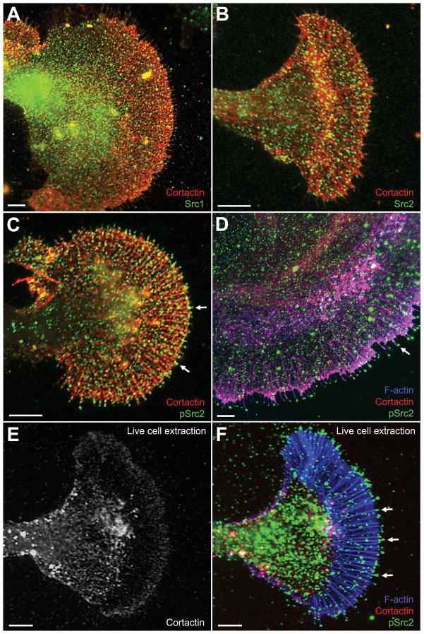
Figure 4. Cortactin and Src colocalization in growth cones.
Cultured Aplysia bag cells were fixed, extracted, and double labeled for cortactin together with Src1 (A), Src2 (B), activated Src2 (pSrc2; C), or triple labeled for cortactin together with F-actin and activated Src2 (D). Regions in magenta denote the colocalization of F-actin and cortactin. Note the enriched pSrc2 staining at the tip of filopodia when compared to total Src2 (arrows in C and D). E–F: Triple staining for cortactin, F-actin, and pSrc2 after live cell extraction. Cortactin was strongly removed from actin bundles in the P domain, but remained in the ruffling zone (E). Activated Src2, on the other hand, remained at the tip of filopodia (arrows in F), which were identified by F-actin staining. The pSrc2 staining outside the growth cone (mostly at the top of image F) is due to cell debris accumulated during the plasma membrane removal. Bars: 10 μm.

