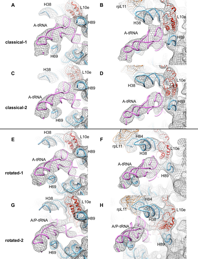
Figure 4. Comparison of A-site tRNA contacts with 60S ribosomal subunit for all four sub-populations described.
(from top to the bottom: A, B, classical-1; C, D, classical-2; E, F, rotated-1; G, H rotated-2). (A, C, E, G) Illustration of different contacts between A-site tRNA and H69. (B, D, F, H) Demonstration of the potentially dynamic interaction between the A-site tRNA elbow and components of the 60S subunit during the transition from classical-1 to rotated-2 intermediate states. Models for rRNA and separated proteins are derived from the X-ray structure of the yeast 80S ribosome ((Ben-Shem et al., 2010) PDB ID 3O58)
See alsoFigure S4. Distinct features of 80S ribosomes in the classical-1 and classical-2 subpopulations.
Movie S1. Spontaneous movement of tRNAs relative to the 60S subunit.
Movie S2. Spontaneous movement of tRNAs relative to the 40S subunit.

