
Figure 2. Decreased inflammation and preservation of myelin content after CDDO-TFEA treatment.
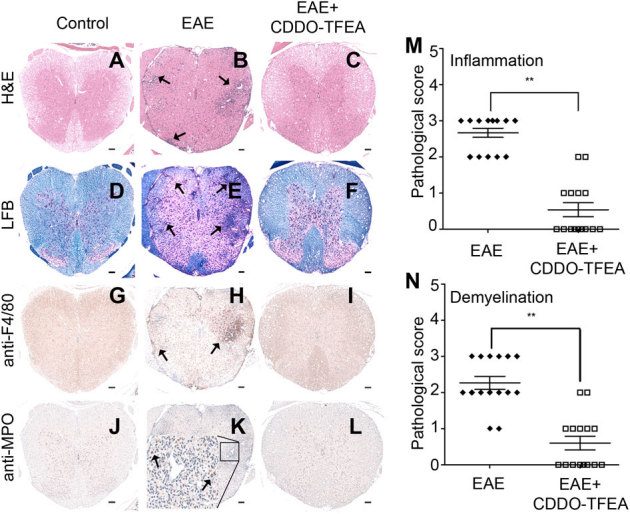
Mice were sacrificed after MOG (35–55) immunization either at the peak of clinical symptoms or at a clinical score of zero and after respective treatments with either vehicle control or CDDO-TFEA and CNS tissues were subjected to histopathological analysis. Representative sections of lumbar spinal cord (A–L) from control mice with clinical signs of EAE or from mice treated with CDDO-TFEA were stained with hematoxilin and eosin to assess inflammation (A–C), with Luxol fast blue to assess myelin content (D–F), with F4/80 to stain macrophages and microglia (G–I) and with myloperoxidase (MPO) to stain neutrophils (J–L). Arrows indicate inflammatory cellular infiltrates (B), demyelinated areas (E) and macrophage and neutrophil infiltrates (H & K). A pathologist blinded to subject identity scored sections taken from each animal for (M) inflammation and (N) demyelination on the scale of 0 to 3 (Scale bars, 100μm). **, P , 0.01, Student's t test.