
Figure 4. CDDO-TFEA inhibits lymphocyte proliferation and modulates iNOS and Hmox-1 protein expression in the CNS of affected mice.
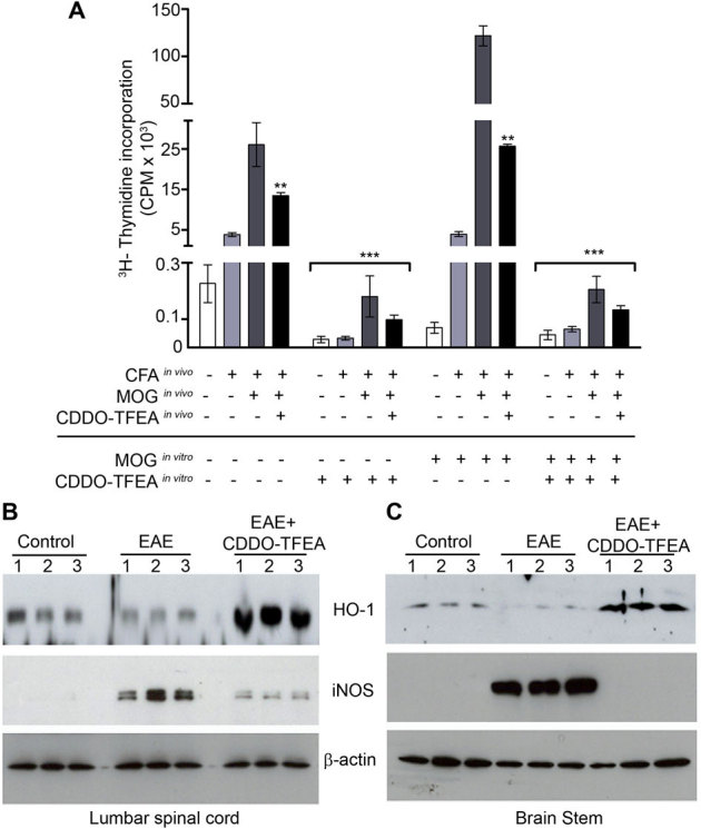
(A) Lymphocytes were collected from control mice, mice with EAE (at clinical score 5) or from mice with EAE following treatment with CDDO-TFEA (at clinical score 0) and cultured for 72 hrs in the presence or absence of MOG (35–55), with or without CDDO-TFEA. During last 16 hrs, 3H-thymidine was added to cultures and the incorporated amount of3H-thymidine in cells was measured as CPM. Protein lysates were prepared from lumbar spinal cord (B) and brain stem (C) of control mice, mice with EAE (at clinical score 5) or mice with EAE following CDDO-TFEA treatment (at clinical score 0) and subjected to Western blot analysis to check iNOS and Hmox-1 protein levels. Equal loading of samples was confirmed by actin immunodetection. Data is presented as the mean ± SEM (n = 6). ***, P , 0.001, **, P , 0.01, Student's t test.