
Figure 6. CDDO-TFEA is neuroprotecive in the LPC-induced model of demyelination.
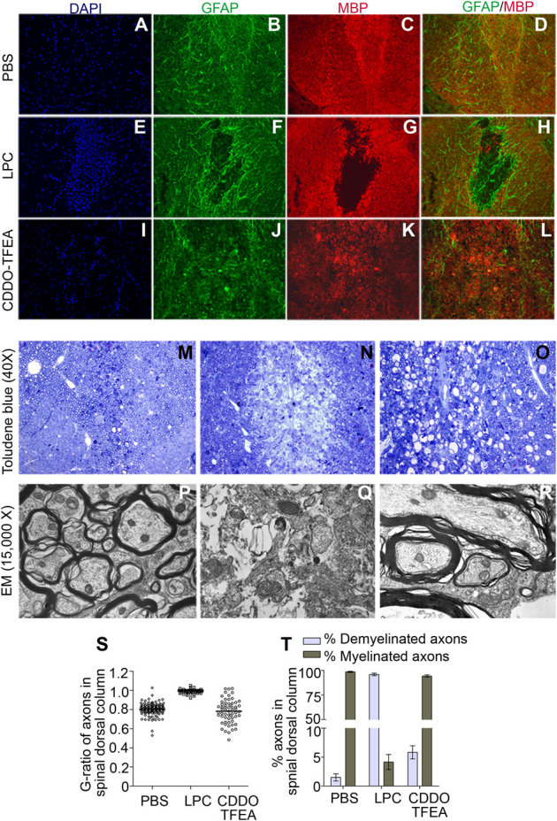
Immunostaining of tissue sections prepared from dorsal columns of the thoracic spinal cord of rats at the site of injection of either PBS (A–D) or LPC (E–H) or LPC+ CDDO-TFEA (I–L). Immunohistochemistry was performed with an anti-MBP antibody (for myelin), anti-GFAP antibody (for astrocytes) and anti-NG2 antibody (for oligodendrocytes). All images are taken at 200X magnification. Tissue sections prepared from spinal cord segments at sites injected either with LPC or LPC followed by CDDO-TFEA were subjected to toludene blue staining (M–O) or electron microscopy (P–R) to analyze the myelin content on neurons at the site of each lesion. G-ratios of the axons in dorsal columns (S) and quantification of the percentage of demyelinated and myelinated axons in dorsal columns of spinal cord at the site of LPC injection (T) are shown here.