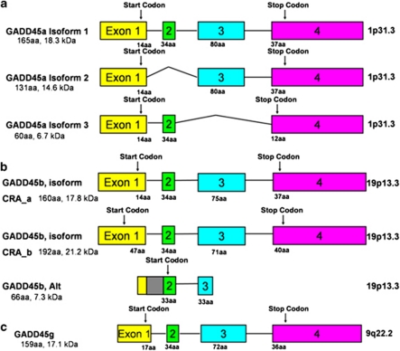
Figure 2.
Exon–intron structure and alternative transcripts of human GADD45 genes. Exons are shown as boxes and introns are shown as lines. Despite the three GADD45 genes being located on different chromosomes there are substantial similarities in terms of gene structure and protein composition. All three proteins are 17–18 kDa in mass and highly acidic. They share 56% protein sequence identity. In (a), are the three known splice variants of GADD45a. Isoform 1 contains four exons, while the other two splice variants are missing the second or third exon respectively. In (b), are the three splice variants of GADD45b. Isoform CRA_a and CRA_b differ based on their start sites. Another splice variant, referred to here as ‘GADD45b, Alt' includes the first intron (gray box), and translation begins at the second exon. This isoform is noted on the NCBI database (AAT38867.1) and in (Ying et al, 2005). In (c), is the gene structure of GADD45g.

