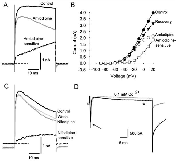
Fig. 4.
Evidence for an L-type Ca2+ current in calyx terminals. A) Control current, current after application of 7 μM amlodipine and the amlodipine-sensitive current obtained by subtraction (dashed line) are shown in a P30 calyx in response to a depolarizing voltage step to +20 mV. Zero-current level is indicated by thin dashed line. B) The I-V plot shows steady-state currents at a series of voltage steps for the same cell as in A. Control currents (filled circles) currents in 7 μM amlodipine (open circles), the amlodipine-sensitive current obtained by subtraction (open squares) and currents following drug washout (filled triangles) are shown. C) Nifedipine application reduced outward current in a P33 calyx. Control current, current in the presence of 20 μM nifedipine (dark grey line) and a partial recovery following washout (light grey line) are shown in response to a +21 mV depolarizing voltage step. The nifedipine-sensitive current is shown by the dashed line. Zero-current level is indicated by the thin dashed line. D) Effect of Cd2+ on slow inward currents in a calyx terminal (P28). Electrode solution contained Cs+ in place of K+. Superimposed currents following a voltage step to −10 mV are shown and include a rapid transient INa (arrow) followed by a slowly activating inward current (*). The slow inward current and inward tail current after the test step were greatly reduced in the presence of 100 μM CdCl2 (grey current trace), indicating the presence of Ca2+ current

