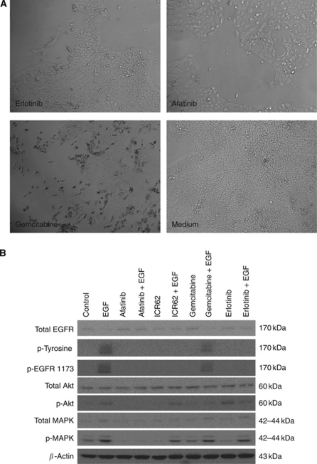
Figure 2.
(A) Morphology of BxPC-3 cells following growth inhibitory concentrations of erlotinib, afatinib and gemcitabine (μM) compared with treatment with medium alone (original magnification × 20). (B) Effect of afatinib, erlotinib and ICR62 on EGF-induced phosphorylation of tyrosine, EGFR, MAPK and Akt in BxPC-3 cells. BxPC-3 cells were cultured to near-confluency in growth medium containing 10% FBS, then treated in 0.1% FBS medium containing 400 nM of TKI, mAb ICR62 (400 nM) or gemcitabine (100 nM) for 24 h at 37 °C. Following incubation with the inhibitors, cells were stimulated with 20 nM of EGF for 15 min. Then, treated cells were lysed, protein samples were separated by SDS–PAGE, transferred onto PVDF membranes, an probed with antibodies specific for the molecule of interest.

