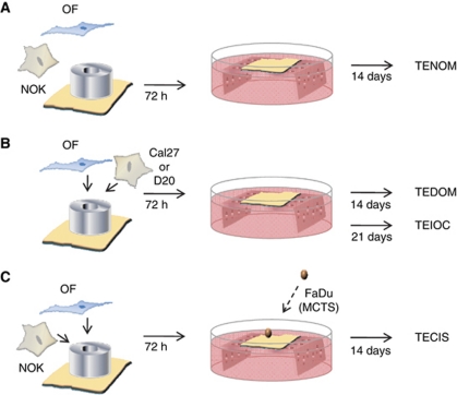
Figure 1.
Schematic illustration showing the methodology for producing the full-thickness tissue-engineered oral mucosal models. To produce the tissue-engineered normal oral mucosa model (TENOM), normal oral fibroblasts (OFs) and normal oral keratinocytes (NOKs) were seeded onto a DED scaffold within a 0.8 cm2 steel ring. After 72 h, the composites were raised to an air/liquid interface and cultured for a further 14 days (A). To recreate an in vitro model of dysplastic oral mucosa (TEDOM) and early invasive oral carcinoma (TEIOC), the NOKs were replaced with OSCC cells (Cal27 or D20) and cultured for either 14 or 21 days, respectively (B). The addition of a MCTS to the TENOM produced a model closely mimicking carcinoma in situ (TECIS) (C).

