Figure 2.
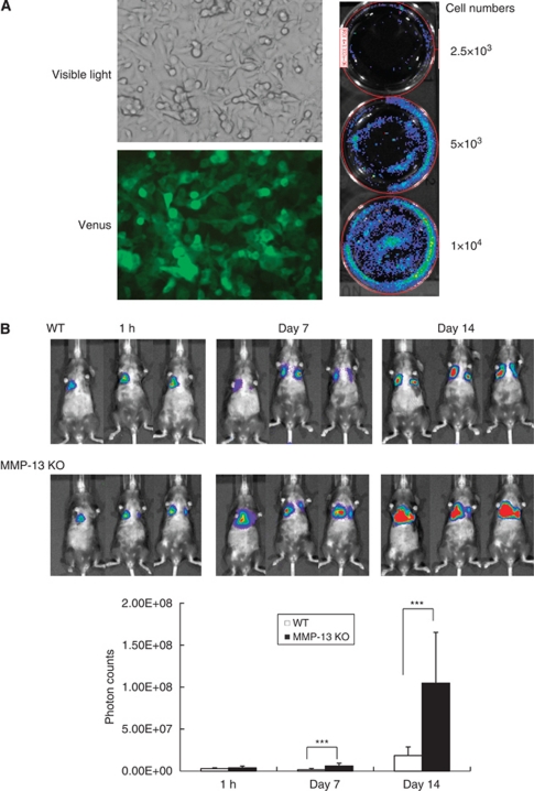
Establishment of B16BL6Venus−Luc melanoma cells and monitoring of lung metastases after the intravenous injection into WT and MMP-13 KO mice. (A) Establishment of B16BL6Venus−Luc melanoma cells. The cells show positive staining under fluorescence microscopy for Venus, and photon counts are dependent on the cell number after addition of luciferin to culture media of the cells. (B) Time-course changes of lung metastases after intravenous injection of B16BL6Venus−Luc cells. Photon counts of whole lungs were counted at 1 h, 7 days and 14 days after intravenous injection of 5 × 10⁴ cells into WT and MMP-13 KO mice (n=5 per group) by using LIVING IMAGE 3.0 software. Bars, mean±s.d. ***P<0.001.

