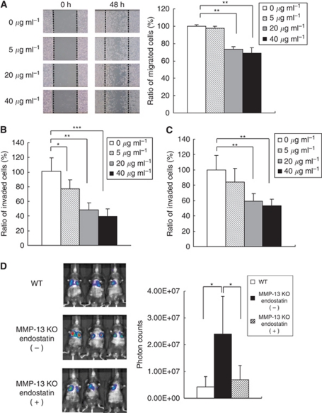
Figure 5.
Inhibitory effects of endostatin on the migration, Matrigel invasion, transendothelial invasion and lung metastases of B16BL6 melanoma cells. (A) Effects of endostatin on the migration of the melanoma cells. B16BL6 melanoma cells in monolayer culture were wounded and migration activity was monitored at 24 and 48 h in the presence of endostatin (0, 5, 20 or 40 μg ml−1) in culture medium containing 5 mM hydroxyurea. Migration areas of the melanoma cells were determined by measuring cell migration areas using Image J software. Results are expressed as percentage of migration area compared with control (0 μg ml−1). Bars, mean±s.d. **P<0.01. (B) Effects of endostatin on Matrigel invasion of B16BL6 melanoma cells. Melanoma cells were added to the upper compartment of Matrigel invasion chamber and number of the cells that invaded Matrigel matrix membrane was counted as described in Materials and Methods. Percentage of invaded cells compared with control (0 μg ml−1) is shown. Bars, mean±s.d. *P<0.05; **P<0.01; ***P<0.001. (C) Effects of endostatin on transendothelial invasion of B16BL6 melanoma cells. Melanoma cells were added to the upper compartment of Matrigel invasion chamber that had been covered with an endothelial cell layer and number of the cells that invaded the endothelial cell layer and Matrigel membrane was counted. Percentage of invaded cells compared with control (0 μg ml−1) is shown. Bars, mean±s.d. **P<0.01. (D) Inhibition of lung metastases by restoration of endostatin in MMP-13 KO mice. Endostatin was intraperitoneally injected into MMP-13 KO mice on days 1, 2, 3 and 4 after intravenous injection of B16BL6Venus−Luc cells and lung metastases were measured by bioluminescence imaging at 2 weeks after the injection. Photon counts were compared between WT mice and MMP-13 KO mice with or without endostatin administration. *P<0.05.

