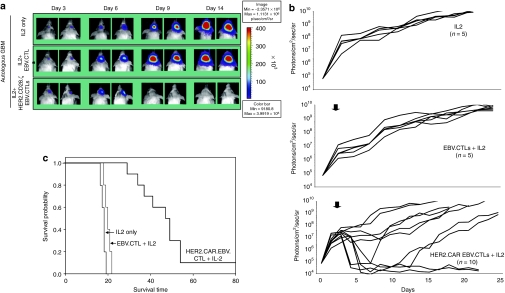
Figure 8.
Adoptively transferred HER2-CTLs induce regression of HER2-positive xenografts in vivo. 5 × 10⁴ eGFP.FFLuc-expressing HT-1080 cells were injected stereotactically into the caudate nucleus of 9–12 week old SCID mice followed by intratumoral injection of 2 × 10⁶ HER2-CTLs or NT EBV-CTLs on day 6 after tumor inoculation. (a) Tumors grew progressively in untreated mice as shown for two representative animals (upper row) and in mice receiving NT EBV-CTLs (middle row), while tumors regressed over a period of 2–5 days in response to a single injection of HER2-CTLs generated from the same donor (lower row). (b) Quantitative bioluminescence imaging: HER2-CTLs induced tumor regression when compared to NT EBV-CTLs (two-tailed P value = 0.006, Mann–Whitney U test). Solid arrows: time of T-cell injection; open arrows: background luminescence (mean~105 photon/sec/cm2/sr); n, number of animals tested in each group. (c) Kaplan–Meier survival curve: Survival analysis performed 80 days after tumor establishment. Mice treated with HER2-CTLs had a significantly longer survival probability (P < 0.007) in comparison to untreated mice and mice that received NT EBV-CTLs. CTLs, cytotoxic T lymphocytes; HER2, human epidermal growth factor receptor 2; EPV, Epstein-Barr virus; NT, nontransfected.

