Figure 4.
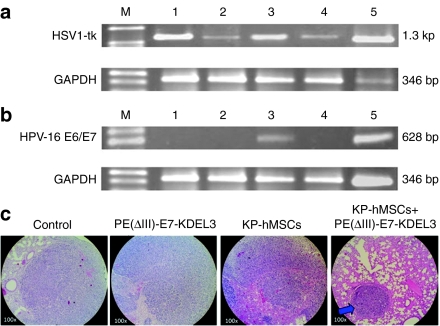
Ex vivo validation of mesenchymal stem cells (MSCs) targeting and vaccine/MSCs combined treatment in pulmonary metastatic tumor. (a) The presence of HSV1-tk gene in the lung tissues from different treatment groups was detected using reverse transcription (RT)-PCR. There were five different groups namely, without any treatment (lane 1), vaccine only treatment (lane 2), MSCs only treatment (lane 3), the combined treatment (lane 4), and positive control NG4TL4-TK cells (lane 5). HSV1-tk gene was used as an indicator for that the pulmonary tumors were developed by NG4TL4-TK cells, and could be detected in all groups. (b) RT-PCR detection of HPV-16 E6/E7 expression in lung tissues from the mice received different treatments. The group arrangement is the same as in a except lane 5, which represents a positive control, MSCs only. HPV-16 E6/E7 gene in the MSC was detected in MSCs only treatment group (lane 3) and MSCs control (lane 5). GAPDH was used as an internal standard. M represents DNA ladder. (c) Hematoxylin and eosin (H&E) stained parafilm sections of the lung tissues from different treatment groups. Pulmonary alveoli were appeared more intact in the combined-treatment group than in other groups. Blue arrow indicated tumor nodule formation.

